Findings make a strong case for harnessing the power of artificial intelligence in CT imaging
June 10, 2019
TROY, N.Y. —Machine learning has the potential to vastly advance medical imaging, particularly computerized tomography (CT) scanning, by reducing radiation exposure and improving image quality.
Those new research findings were just published in Nature Machine Intelligence by engineers at Rensselaer Polytechnic Institute and radiologists at Massachusetts General Hospital and Harvard Medical School.
According to the research team, the results published in this high-impact journal make a strong case for harnessing the power of artificial intelligence to improve low-dose CT scans.
“Radiation dose has been a significant issue for patients undergoing CT scans. Our machine learning technique is superior, or, at the very least, comparable, to the iterative techniques used in this study for enabling low-radiation dose CT,” said Ge Wang, the Clark & Crossan Endowed Chair Professor of biomedical engineering at Rensselaer, and a corresponding author on this paper. “It’s a high-level conclusion that carries a powerful message. It’s time for machine learning to rapidly take off and, hopefully, take over.”
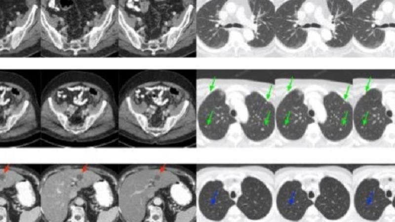
Low-dose CT imaging techniques have been a significant focus over the past several years in an effort to alleviate concerns about patient exposure to X-ray radiation associated with widely used CT scans. However, decreasing radiation can decrease image quality.
To solve that, engineers worldwide have designed iterative reconstruction techniques to help sift through and remove interferences from CT images. The problem, Wang said, is that those algorithms sometimes remove useful information or falsely alter the image.
The team set out to address this persistent challenge using a machine learning framework. Specifically, they developed a dedicated deep neural network and compared their best results to the best of what three major commercial CT scanners could produce with iterative reconstruction techniques.
This work was performed in close collaboration with Dr. Mannudeep Kalra, a professor of radiology at Massachusetts General Hospital and Harvard Medical School, who was also a corresponding author on the paper.
The researchers were looking to determine how the performance of their deep learning approach compared to the selected representative iterative algorithms currently being used clinically.
Several radiologists from Massachusetts General Hospital and Harvard Medical School assessed all of the CT images. The deep learning algorithms developed by the Rensselaer team performed as well as, or better than, those current iterative techniques in an overwhelming majority of cases, Wang said.
Researchers found that their deep learning method is also much quicker, and allows the radiologists to fine-tune the images according to clinical requirements, Dr. Kalra said.
These positive results were realized without access to the original, or raw, data from all the CT scanners. Wang pointed out that if original CT data is made available, a more specialized deep learning algorithm should perform even better.
“This has radiologists in the loop,” Wang said. “In other words, this means that we can integrate machine intelligence and human intelligence together in the deep learning framework, facilitating clinical translation.”
He said that these results confirm that deep learning could help produce safer, more accurate CT images while also running more rapidly than iterative algorithms.
“We are excited to show the community that machine learning methods are potentially better than the traditional methods,” said Wang. “It sends the scientific community a strong signal. We should go for machine learning.”
This research by Wang’s team is among the significant advancements consistently being made by faculty in the Biomedical Imaging Center within the Center for Biotechnology and Interdisciplinary Studies (CBIS) at Rensselaer.
“Professor Wang’s work is an excellent example of how advances in artificial intelligence, and machine and deep learning, can improve biomedical tools and practices by addressing hard problems—in this case helping to provide high-quality CT images using a lower radiation dose. Transformative developments from these collaborative teams will lead to more precise and personalized medicine,” said Deepak Vashishth, director of CBIS.
Hongming Shan, a postdoctoral researcher at Rensselaer, is the first author of the paper. Uwe Kruger, professor of practice in biomedical engineering at Rensselaer, was instrumental when it came to statistical analysis in this project. Radiologists from Massachusetts General Hospital in Boston and Ramathibodi Hospital in Bangkok are also coauthors on this research. This work was supported in part by a grant from the National Institute of Biomedical Imaging and Bioengineering within the National Institutes of Health.