Engineering Researchers at Rensselaer Polytechnic Institute Develop “Phantoms” To Make Medical Imaging Safer for Overweight Individuals
April 12, 2012
Most medical imaging equipment is not designed with overweight and obese patients in mind. As a result, these individuals can be exposed to higher levels of radiation during routine X-ray and CT scans.
A new study from Rensselaer Polytechnic Institute is the first to calculate exactly how much additional radiation obese patients receive from a CT scan. Research results show the internal organs of obese men receive 62 percent more radiation during a CT scan than those of normal weight men. For obese women, it was an increase of 59 percent.
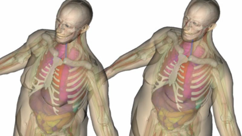
New technology developed at Rensselaer by nuclear engineering expert X. George Xu could help solve this problem. Xu’s research team created ultra-realistic 3-D computer models of overweight and obese men and women, and used computer simulations to determine how X-rays interact with the different body types. These models, known as “phantoms,” can help empower physicians to configure and optimize CT scanning devices in such a way that minimizes how much radiation a patient receives.
“Radiation exposure is cumulative over a patient’s lifetime. The risk associated with a radiation dose from a single CT scan is relatively small when compared with the clinical benefit of the procedure. But patients are increasingly undergoing multiple CT scans and other radiation-based procedures, which can lead to unnecessary radiation risk. Regretfully, our study shows that obese and overweight patients can be exposed to an even greater level of radiation,” said Xu, head of the Nuclear Engineering Program and a professor in the Department of Mechanical, Aerospace, and Nuclear Engineering (MANE) at Rensselaer. “Our new study brings us one step closer to minimizing radiation exposure and mitigating this risk to patients.”
Results of the Xu’s study were published today in the journal Physics in Medicine & Biology. The study may be viewed online at: http://m.iopscience.iop.org/0031-9155/57/9/2441.
Currently, if technicians use normal equipment settings to perform a CT scan on an obese patient, the resulting images are blurry as the X-ray photons have to travel further and make their way through layers of fat. As a result, technicians generally adjust the equipment to a more powerful setting, which produces a better image but exposes the obese patient to additional radiation. There is no mechanism for discerning the setting levels that will provide an optimal balance of the highest image clarity and the lowest radiation dose.
These new phantoms for overweight and obese patients will be part of a forthcoming software package, VirtualDose, developed by Xu and his team. VirtualDose aims to enable the creation of a personalized, ultra-realistic phantom of any patient undergoing a CT scan. The program takes into consideration a patient’s individual characteristics, including age, sex, height, weight, and even if a woman is pregnant. By entering these data into the software, VirtualDose quickly creates a phantom that accurately models the patient’s internal organs. These phantoms will allow physicians and researchers to compare the radiation doses a patient will get from different CT scanner settings, and then choose the most appropriate configuration.
VirtualDose will also enable physicians to keep a highly accurate record of how much radiation patients are exposed to over their lifetime. California recently became the first state in the United States to require radiation dose records for patients undergoing CT examinations.
Along with Xu, authors of the study are research associate Aiping Ding; graduate students Matthew Mille and Tianyu Liu; and lecturer and campus radiation safety officer Peter Caracappa, all of the Nuclear Engineering Program and MANE at Rensselaer.
The research is funded primarily by the National Institutes of Health (NIH), with additional support from the National Institute of Biomedical Imaging and Bioengineering, the U.S. Department of Energy (DOE) Office of Nuclear Energy’s Nuclear Energy University Programs (NEUP), and the Health Physics Society.
The Nuclear Engineering Program at Rensselaer is among the oldest in the nation, dating back to the late 1950s when the university built an electron accelerator.
For more information on Xu’s research at Rensselaer, visit:
- Wall Street Journal: Scientists Find Safer Ways To Test Medical Procedures
http://on.wsj.com/HayzNv - Patient Safety: Reducing the Risks of Radiation Exposure From CT Scans and X-Rays
http://news.rpi.edu/update.do?artcenterkey=2876 - Safer, More Accurate Radiation Therapy for Expecting Mothers
http://news.rpi.edu/update.do?artcenterkey=2377 - The Phantom Patient
http://approach.rpi.edu/2011/12/20/the-phantom-patient-2/ - A Virtual Patient To Simulate Real-Time Organ Motions
http://www.rpi.edu/research/magazine/summer07/virtual_patient1.html - Rensselaer Radiation Measurement and Dosimetry Group
http://www.rpi.edu/dept/radsafe/public_html/index.htm - Nuclear Engineering Program
http://www.rpi.edu/dept/ne