Deep Neural Network developed at Rensselaer could improve cancer care
November 13, 2019

TROY, N.Y. — Improving the detection, diagnosis, and treatment of diseases like cancer will require more detailed, rapid, and agile imaging technology that can show doctors not just what a specific organ looks like, but also what’s happening within the cells that make up those tissues.
In research published in Proceedings of the National Academy of Sciences, a team from Rensselaer Polytechnic Institute developed and demonstrated a new technique for fluorescence lifetime imaging of tissue and cells in a fast and comprehensive manner — laying the groundwork for use in a clinical setting.
“We are providing tools that are going to be far more amenable for the end users, meaning the biologists, but also the surgeon,” said Xavier Intes, a professor of biomedical engineering who led this research for Rensselaer.
Fluorescence lifetime imaging has long been a helpful way for scientists and engineers to see molecular-level interactions within cells — a necessary tool when trying to identify cancer and other diseases, and evaluate the effectiveness of drugs.
Traditionally, Intes said, producing an image this way has required a lot of time and complex mathematical tools that rely heavily on the user, which makes it hard to produce consistent and reproducible images. Those difficulties have been barriers to using this type of imaging in a clinical setting.
To overcome those challenges, the Rensselaer team designed a deep neural network (DNN) to automatically set the mathematical parameters that a human typically would, while also producing a detailed image that can show interactions in cells or tissue as they’re happening.
This work builds on the Rensselaer team’s previous research, where the reserachers developed a method for quickly reconstructing a single lifetime image. This new approach reconstructs multiple lifetime images at the same time, providing a comprehensive view of multiple biological processes happening within the tissue and cells, said Pingkun Yan, an assistant professor of biomedical engineering and member of the Center for Biotechnology and Interdisciplinary Studies, who also worked on this research.
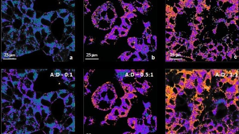
The team, in partnership with biologists at Albany Medical College, tested this new technique by imaging cancer cells under the microscope and in living systems. What they observed, Intes said, was that their DNN performed as well as, or in some cases better than, commercial software currently being used. The team also found this technique required less light while still producing detailed images, which is critical for biological applications.
The researchers’ success brings the field closer to being able to use fluorescence lifetime imaging in a clinical setting in order to evaluate the effectiveness of a particular drug on a person’s individual cancer cells — a key tool needed to enable precision medicine.
The researchers also were able to apply this DNN to visualize the level of activity within cells, a process known as metabolic imaging. This approach could help guide surgeons in the operating room as they work to identify which tissue is healthy, and which is diseased and should be removed.
“This is an enabling technology for many clinical applications. For instance, it may be used for in vivo real-time imaging of a tumor, which may help surgeons see the lesion during their procedures, enabling them to completely remove cancer tissue with minimal damage to healthy tissue,” Yan said.
The team is currently working with Dr. Brett Miles from the Icahn School of Medicine at Mount Sinai to implement this approach for head and neck cancer surgeries.
This work was supported by three grants from the National Institutes of Health.