New technology will give researchers a deeper look at cancer and effectiveness of treatments
September 18, 2019
TROY, N.Y. — If researchers could observe drug delivery and its effect on cancer cells in real time, they would be able to tailor treatment options with unprecedented specificity.
An academic-industrial partnership between engineers at Rensselaer Polytechnic Institute, molecular and cellular biologists at Albany Medical College, and engineers at MARS Bioimaging Ltd aims to make this a reality for the treatment of breast cancer through the combination of highly innovative X-ray and optical imaging technologies.
“We are combining cutting-edge technologies that we had to develop ourselves. There is no synergy like this in the world at this point,” said Xavier Intes, a professor of biomedical engineering at Rensselaer. “We are uniquely positioned to develop the next imaging systems to support precision medicine.”
This multidisciplinary project is being supported for five years by a nearly $2.9 million grant from the National Institutes of Health (NIH). In a highly competitive field, NIH rated it among the top 1% of research proposals submitted for funding.
Intes and Ge Wang, co-director and director of the Biomedical Imaging Center within the Center for Biotechnology and Interdisciplinary Studies, are leading this research for Rensselaer with Margarida Barroso, a professor in the Department of Molecular and Cellular Physiology at Albany Medical College. The team will also be joined by Anthony Butler, a radiologist with MARS Bioimaging Ltd. in New Zealand and a specialist in the X-ray spectral photon-counting imaging technology that will be used in this research.
“This work on breast cancer will pave the way toward a better diagnosis and management for a broad, wide range of cancers,” Butler said.
The Rensselaer engineers will combine X-ray photon-counting and optical time-resolving imaging technologies that their teams have developed, enabling scientists to watch cancer in living systems as it reacts to treatment. Barroso and her team will develop and characterize how different types of breast tumors are able to take in clinical drugs, used in this research, as imaging probes to visualize their location in tumors.
Wang and his team, in collaboration with MARS Bioimaging, have developed a photon-counting X-ray method for diffuse optical tomography that uses information gathered from the energy of each and every individual photon to generate an image — in color — that differentiates between different materials like soft tissue, bone, water, contrast agents, and pharmaceuticals. This complements the functional and molecular properties that optical imaging reveals about each organ and tissue.
Intes and his research group, in collaboration with Albany Medical College, have developed novel optical imaging methods that allow researchers to observe — down to the cellular and molecular level — delivery of drugs to the cell, and any interaction that might happen.
Combining the two technologies will help researchers single out the cancer cells and monitor their responses to therapeutic agents.
“X-ray and optical are highly synergistic modalities working together. This is a perfect marriage,” Wang said.
Both teams are also developing the next generation of imaging algorithms using deep learning methodologies to produce clearer images than were previously possible.
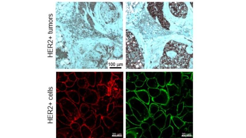
In this specific project, the team will focus on a type of breast cancer that expresses human epidermal growth factor receptor 2, or HER2. Targeted therapies for this kind of cancer have demonstrated great promise in clinical settings, the researchers said, but drug resistance over time remains a problem.
“Numerous drugs have been developed recently to target breast cancer, but their ability to reach all cancer cells in a tumor is still lacking,” Barroso said. “This imaging drug-delivery study will allow us to understand what prevents drugs from reaching different areas of the tumor and, thus, how to act to improve drug delivery to combat breast cancer.”
This new integrated imaging approach will allow the biologist team at Albany Medical College to explore why that resistance is happening by enabling scientists to understand specific cancer mechanisms more deeply, assess in real time if a drug is accurately and thoroughly being delivered to the tumor cells, and if the drug is effective and persistent.
“Cancer is not static. Cancer is dynamic,” Intes said. “When you start to introduce drugs, it can change rapidly and even completely, so noninvasive imaging is important.”