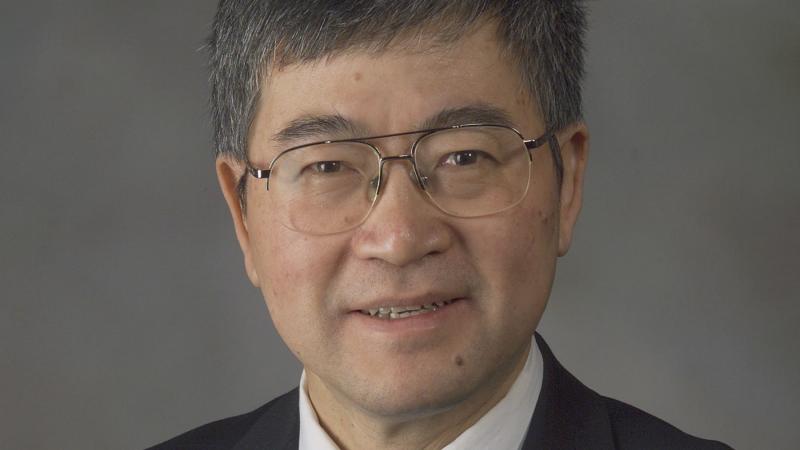
Rensselaer researcher has contributed significantly to advancing biomedical imaging
December 3, 2019

TROY, N.Y. — Ge Wang, the Clark and Crossan Endowed Chair of biomedical engineering and director of the Biomedical Imaging Center at Rensselaer Polytechnic Institute, has been named a fellow of the National Academy of Inventors (NAI).
Election to NAI fellow is the highest professional distinction given to academic inventors. It is bestowed on those who have created or facilitated inventions that have improved quality of life, economic development, and the welfare of society.
“Professor Wang has made exceptional contributions in the area of biomedical imaging,” said Deepak Vashishth, director of the Center for Biotechnology and Interdisciplinary Studies, of which the Biomedical Imaging Center is an important part. “The methods he has developed are necessary tools in vastly improving human health and eventually moving toward personalized medicine.”
Throughout his distinguished career, Wang has focused on developing approaches to improve medical images. The methods that he has created — related to CT imaging and other forms of tomography — aim to deepen understanding of diseases and improve diagnosis and treatment.
He published the first helical/spiral cone-beam/multi-slice CT algorithm in 1991, followed by more than a hundred papers in this area. This type of CT is now used in hospitals worldwide.
“Dr. Wang is a pioneer. He is applying advanced data, artificial intelligence, and computing techniques to provide a much clearer look into the human body,” said Shekhar Garde, dean of the School of Engineering. “These ‘new eyes’ will enable faster diagnosis of complex diseases and more effective treatments.”
Wang and his team are currently working on a cross-disciplinary study, supported by the National Institutes of Health, to combine highly innovative photon-counting X-ray and lifetime optical imaging technologies — developed at Rensselaer — to allow biologists to observe drug delivery and its effect on cancer cells, in vivo and in real time.
Earlier this year, in a paper published in Nature Machine Intelligence, Wang and his collaborators showed that machine learning can improve low-dose CT imaging by reducing radiation exposure without compromising image quality.
In addition to research, Wang is also interested in innovative pedagogy. His TED-Ed lecture on How X-rays See through Your Skin has received more than one million views.
Wang has published more than 480 journal articles, and is a fellow of a number of other professional associations including the Institute of Electrical and Electronics Engineers, the Optical Society of America, the American Association of Physics in Medicine, the American Institute for Medical and Biological Engineering, and the American Association for the Advancement of Science.
His lab is funded by several companies targeting clinical translation of machine learning, especially deep learning techniques. He and his co-authors are in the process of publishing the book Machine Learning for Tomographic Imaging through IOP Publishing.
He joins a distinguished group of fellow academic inventors. The fellows in the 2019 class represent 136 research universities and research institutes worldwide, and collectively hold more than 3,500 issued U.S. patents.
The NAI Fellows Induction Ceremony will be held on April 10, 2020, in Phoenix, Arizona