To Improve Surgeons’ Skills, Researchers Will Tap Directly Into Their Brains

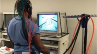
Researchers at Rensselaer Polytechnic Institute envision a day when surgeons will benefit from personalized training, rather than sheer practice repetition, thanks to novel neuroimaging and artificial intelligence methodologies. Under this method, surgeons would complete technical tasks while images of their brain activity reveal how well they have mastered critical skills.
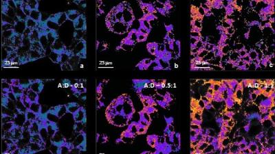
New Imaging Technique Enables the Study of 3D Printed Brain Tumors

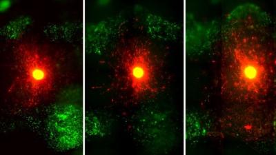
TROY, N.Y. — Glioblastomas are complex, fast-growing malignant brain tumors that are made up of various types of cells. Even with aggressive treatment — which often includes surgery, radiation, and chemotherapy — glioblastomas are difficult to treat, leading to an average survival of 11-15 months.

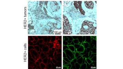
New Technique Aims to Improve Imaging of Cells

TROY, N.Y. — Improving the detection, diagnosis, and treatment of diseases like cancer will require more detailed, rapid, and agile imaging technology that can show doctors not just what a specific organ looks like, but also what’s happening within the cells that make up those tissues.

Integrated Imaging Technology Aims To Provide Real-Time Look at Cancer Treatment

TROY, N.Y. — If researchers could observe drug delivery and its effect on cancer cells in real time, they would be able to tailor treatment options with unprecedented specificity. An academic-industrial partnership between engineers at Rensselaer Polytechnic Institute, molecular and cellular biologists at Albany Medical College, and engineers at MARS Bioimaging Ltd aims to make this a reality for the treatment of breast cancer through the combination of highly innovative X-ray and optical imaging technologies.

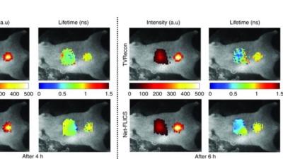
Improving Molecular Imaging using a Deep Learning Approach

TROY, N.Y. — Generating comprehensive molecular images of organs and tumors in living organisms can be performed at ultra-fast speed using a new deep learning approach to image reconstruction developed by researchers at Rensselaer Polytechnic Institute. The research team’s new technique has the potential to vastly improve the quality and speed of imaging in live subjects and was the focus of an article recently published in Light: Science and Applications, a Nature journal.

Advancing Personalized Anti-Cancer Drugs

Researchers at Rensselaer Polytechnic Institute and Albany Medical Center are working together to develop three-dimensional bioprinting and imaging techniques that will generate and analyze tumor models in the laboratory, with the goal of accelerating the development and optimization of personalized anti-cancer drugs.

New Bioimaging Technique Is Fast and Economical

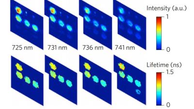
A new approach to optical imaging makes it possible to quickly and economically monitor multiple molecular interactions in a large area of living tissue – such as an organ or a small animal; technology that could have applications in medical diagnosis, guided surgery, or pre-clinical drug testing.

Back to top